Upper Leg Tendon Anatomy : Concept Conceptual 3D Image & Photo (Free Trial) | Bigstock - Try this movement out by standing on one foot with the other leg.. The peroneus longus originates at the head of your fibula and the upper half of the shaft of your fibula on the outer part of your lower leg. Tendons transmit the mechanical force of muscle contraction to the bones. Upper leg anatomy and function. The axilla and the deltoid region in axial and coronal and axial. There are four muscles in the anterior compartment of the leg.
If you tear your biceps tendon at the shoulder, you may lose some strength in your arm and have pain when you forcefully turn your arm from palm down to palm up. Upper limb trauma programme injuries. Quadriceps tendon to base of patella and onto tibial tuberosity via the patellar ligament action: There are four muscles in the anterior compartment of the leg. The lower leg is comprised of two bones, the tibia and the smaller fibula.
Upper Leg Muscles, Posterior from www.purposegames.com Learn vocabulary, terms and more with flashcards, games and other study tools. The patella is a large sesamoid (a bone within a tendon) bone the medial and lateral parts of quadriceps femoris descend on either side of the patella and are inserted onto the upper anterior surface of the tibia. Tendons are fibrous cords attached to muscles and bone. The lower leg is comprised of two bones, the tibia and the smaller fibula. Find stockbilleder af concept 3d human upper leg anatomy i hd og millionvis af andre royaltyfri stockbilleder, illustrationer og vektorer i shutterstocks samling. The thigh bone, or femur, is the large upper leg bone that connects the lower leg bones (knee joint) to the pelvic bone (hip joint). Together, the upper and lower legs and the feet make up half the length of the human figure. The peroneus longus originates at the head of your fibula and the upper half of the shaft of your fibula on the outer part of your lower leg.
The image is available for download in high resolution quality up to 2946x2946.
The tendons that control movement in your hands, wrists and fingers run through your forearm. Muscles attachment , anatomy atlas. See the pictures and anatomy description of knee joint bones, cartilage, ligaments, muscle and tendons with resources for knee problems & injuries. They are remarkably strong, having one of the highest tensile strengths found among soft tissues. The thigh bone, or femur, is the large upper leg bone that connects the lower leg bones (knee joint) to the pelvic bone (hip joint). Find stockbilleder af concept 3d human upper leg anatomy i hd og millionvis af andre royaltyfri stockbilleder, illustrationer og vektorer i shutterstocks samling. This mri wrist coronal cross sectional anatomy tool is absolutely free to use. Use the mouse scroll wheel to move the images up and down alternatively use the tiny arrows (>>) on both side of the image to move the images. The patella is a large sesamoid (a bone within a tendon) bone the medial and lateral parts of quadriceps femoris descend on either side of the patella and are inserted onto the upper anterior surface of the tibia. The lower leg is comprised of two bones, the tibia and the smaller fibula. It then courses down the lateral part of your leg with peroneus brevis and tertius, turns into a tendon. The achilles tendon (tendo calcaneus or tendo achillis) is the thickest and strongest tendon in the human body. Tendon, tissue that attaches a muscle to other body parts, usually bones.
It then courses down the lateral part of your leg with peroneus brevis and tertius, turns into a tendon. Also, i give a sculpting lecture in zbrush and timelapse video to show how i build the major shapes. There are four muscles in the anterior compartment of the leg. A tendon is the fibrous tissue that attaches muscle to bone in the human body. Use the mouse scroll wheel to move the images up and down alternatively use the tiny arrows (>>) on both side of the image to move the images.
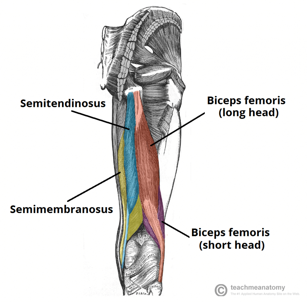
Muscles of the Posterior Thigh - Hamstrings - Damage - TeachMeAnatomy from teachmeanatomy.info Tusindvis af nye billeder af høj kvalitet tilføjes hver dag. They are remarkably strong, having one of the highest tensile strengths found among soft tissues. Iliotibial band syndrome description the iliotibial band is the tendon attachment of hip muscles into the upper leg (tibia) just below the knee to the outer side of the front of the leg. The tendons of the edl can be palpated on the dorsal surface of the foot. Synovial tendon sheaths of right fingers. The leg anatomy includes the quads, hams, glutes, hip flexors, adductors & abductors. Tendinous sheath of right flexor pollicis longus radial bursa. Tendons are fibrous cords attached to muscles and bone.
Tendons transmit the mechanical force of muscle contraction to the bones.
The upper leg begins at the hip and continues down to the knee. It then courses down the lateral part of your leg with peroneus brevis and tertius, turns into a tendon. Movement at the hip joint occurs when you tendons that help you bend or straighten the knee include: The tendons that control movement in your hands, wrists and fingers run through your forearm. Quadriceps tendon to base of patella and onto tibial tuberosity via the patellar ligament action: This mri wrist coronal cross sectional anatomy tool is absolutely free to use. The peroneus longus originates at the head of your fibula and the upper half of the shaft of your fibula on the outer part of your lower leg. Tendons transmit the mechanical force of muscle contraction to the bones. Muscles attachment , anatomy atlas. The human leg, in the general word sense, is the entire lower limb of the human body, including the foot, thigh and even the hip or gluteal region. Upper leg, knee, lower leg, ankle, and foot. Use the mouse scroll wheel to move the images up and down alternatively use the tiny arrows (>>) on both side of the image to move the images. Illustrations of the anatomy of the upper limb.
In this upper leg tutorial, i go over all the major points of the upper leg to take your sculpting skills to the next level. The human leg, in the general word sense, is the entire lower limb of the human body, including the foot, thigh and even the hip or gluteal region. Tendon, tissue that attaches a muscle to other body parts, usually bones. The image is available for download in high resolution quality up to 2946x2946. This mri wrist coronal cross sectional anatomy tool is absolutely free to use.
Quadriceps muscle strain or tear: Torn Quad muscle, Physio Pretoria from physiopretoria.co.za Iliotibial band syndrome description the iliotibial band is the tendon attachment of hip muscles into the upper leg (tibia) just below the knee to the outer side of the front of the leg. Tendinous sheath of right flexor pollicis longus radial bursa. The leg is composed of five distinct sections: The achilles tendon (tendo calcaneus or tendo achillis) is the thickest and strongest tendon in the human body. Upper leg, knee, lower leg, ankle, and foot. To describe the mechanical properties of tendons. Learn vocabulary, terms and more with flashcards, games and other study tools. If you tear your biceps tendon at the shoulder, you may lose some strength in your arm and have pain when you forcefully turn your arm from palm down to palm up.
The tendons of the edl can be palpated on the dorsal surface of the foot.
Iliotibial band syndrome description the iliotibial band is the tendon attachment of hip muscles into the upper leg (tibia) just below the knee to the outer side of the front of the leg. Tendinous sheath of right flexor pollicis longus radial bursa. Peroneal tendonitis affects these tendons, and can make movement difficult and painful. If you tear your biceps tendon at the shoulder, you may lose some strength in your arm and have pain when you forcefully turn your arm from palm down to palm up. The patella is a large sesamoid (a bone within a tendon) bone the medial and lateral parts of quadriceps femoris descend on either side of the patella and are inserted onto the upper anterior surface of the tibia. Artists usually begin their study of the legs by. Tendon, tissue that attaches a muscle to other body parts, usually bones. Legs come in all shapes and sizes, ranging from portly and stout, to the streamlined, almost emaciated legs of runway models, to the muscular legs of athletes. There are four muscles in the anterior compartment of the leg. Upper leg, knee, lower leg, ankle, and foot. Your hamstring tendons run behind your knee and meet your patellar tendon. Quadriceps tendon to base of patella and onto tibial tuberosity via the patellar ligament action: Fibula— a long, thin bone in the lower leg on the lateral side which runs along side the tibia from the knee to the ankle.